
Abstract
Background: Calcaneal fractures are challenging to manage, with minimally invasive techniques aiming to reduce complications. Robotic-assisted surgery offers improved precision, reduced radiation exposure, and better screw placement.
Case presentation: An 85-year-old woman with a Sanders type III intra-articular calcaneal fracture underwent robot-assisted closed reduction and percutaneous screw fixation using the Tirobot II system. The 50-minute procedure achieved accurate alignment and fixation, with rapid pain relief (VAS 10 to 3) and good functional recovery (AOFAS 90) at three months, without complications.
Conclusion: Robot-assisted minimally invasive treatment is effective for calcaneal fractures, providing precise reduction, faster recovery, and minimal complications. Further studies are needed to confirm its long-term benefits.
Keywords: Robot-assisted, Minimally invasive, Calcaneal fracture
Background
Calcaneal fractures, especially intra-articular fractures, are complex to manage and often require surgical intervention to restore joint functionality and anatomical alignment. While traditional open reduction and internal fixation (ORIF) provide excellent fracture visualization, they are associated with significant soft tissue complications, such as infections and delayed healing. In response to these challenges, minimally invasive surgery (MIS) techniques, including percutaneous screw fixation, have gained traction due to their lower complication rates and shorter recovery times [1].
Robot-assisted surgery further enhances the precision and safety of minimally invasive approaches. It enables surgeons to perform complex reductions and screw insertions with greater accuracy and reduced radiation exposure. Research has shown that robot-assisted systems offer superior alignment and screw placement compared to conventional methods, resulting in improved functional outcomes and fewer complications [2]. Robot-assisted surgery also reduces operative time and optimizes postoperative recovery. Studies comparing robot-assisted methods with ORIF indicate that robotic procedures result in fewer wound complications and faster weight-bearing recovery [3]. These benefits align with the principles of minimally invasive surgery: achieving optimal outcomes while minimizing tissue trauma and recovery time. As robotic technology advances, further improvements in surgical precision, efficiency, and patient outcomes are anticipated. Long-term clinical trials and standardized protocols are essential to confirm the full potential of these systems for treating calcaneal fractures and other orthopedic conditions. The goal remains to provide patient-centered care that optimizes function, minimizes complications, and accelerates recovery.
This case report demonstrates how robotic precision can facilitate the accurate insertion of Kirschner wires through small incisions to restore collapsed bone fragments. By integrating robotic guidance, the procedure not only improves reduction accuracy but also minimizes soft tissue trauma, showcasing the potential of robotics in managing complex intra-articular fractures.
Case Presentation
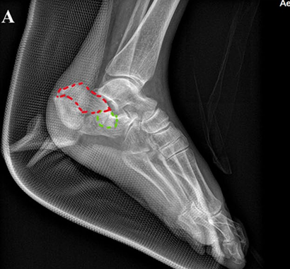
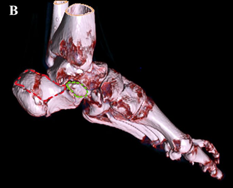
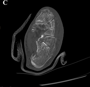
An 85-year-old female presented with left heel pain and inability to bear weight after falling from a height of 2 m. There were no symptoms of headache, dizziness, nausea, vomiting, chest pain, abdominal pain, or back pain. Radiographs and CT scans showed a Sanders type III intra-articular fracture of the right calcaneus with displacement (Fig. 1). Inside the red dotted line is the tongue-type fragment (Fig. 1A). After discussion of treatment options, the patient consented for robot-assisted minimally invasive treatment. The patient reports generally good health with a history of hypertension for 20 years and allergic rhinitis with asthma for 30 years. She has a history of allergy to antipyretic analgesics. She denies any history of infectious diseases, vaccination details are unknown, and she denies any history of trauma, blood transfusions, or blood products.
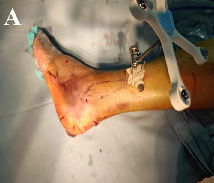
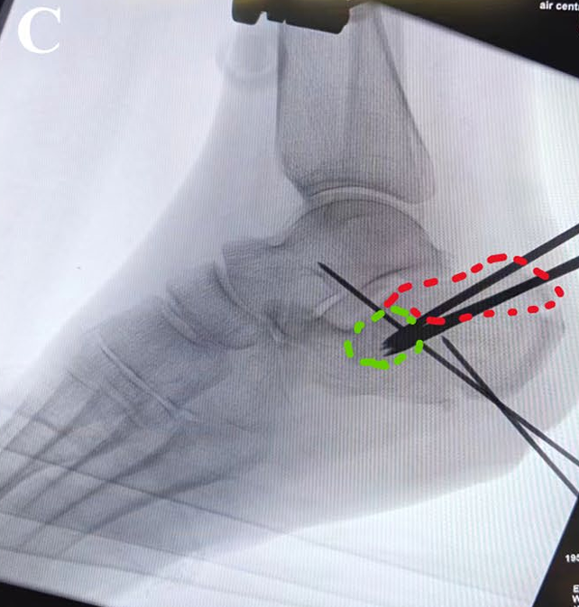
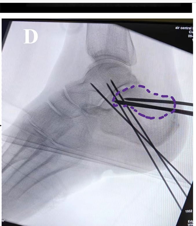
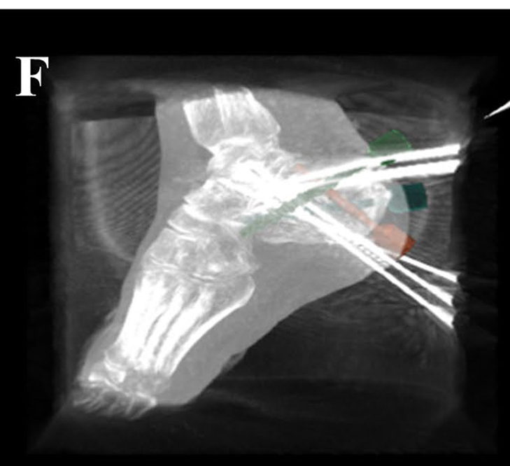
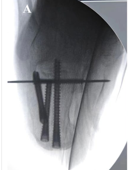
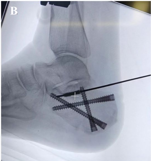
We performed closed reduction and percutaneous screw fixation with assistance of Tirobot II (TINAVI, China). The surgery procedure was as follows. After satisfactory anesthesia, the patient was positioned in the right lateral decubitus position, followed by routine disinfection and draping. The robot tracker was installed at the lower third of the fibula. The calcaneus was scanned with CT, and data were imported into the robot system. Kirschner wires were pre-planned utilizing the robot’s precise positioning. The sophistication of the surgery lies in the use of Steinmann pins for reduction, which need to pass through the tongue-shaped bone fragment (Fig. 2C, fragment 1, red dotted line) to reach the individually fractured bone pieces (Fig. 2C, fragment 2, green dotted line). After penetrating the two fragments, we elevated them as a whole (Fig. 2D, purple dotted line) using the Steinmann pin as joysticks to achieve reduction of the articular surface. Under C-arm fluoroscopy, the fracture alignment and joint surface were deemed satisfactory, with restoration of calcaneal height (from 22 to 40 mm), length (from 59 to 65 mm), Bohler’s angle (from 23 to 26°) and Gissane’s angle (from 65 to 106°). Screws were planned and inserted robotically into specific areas of the calcaneus. The screw positions and lengths were confirmed to be satisfactory under fluoroscopy (Fig. 3, A, B). The whole procedure lasted 50 min.
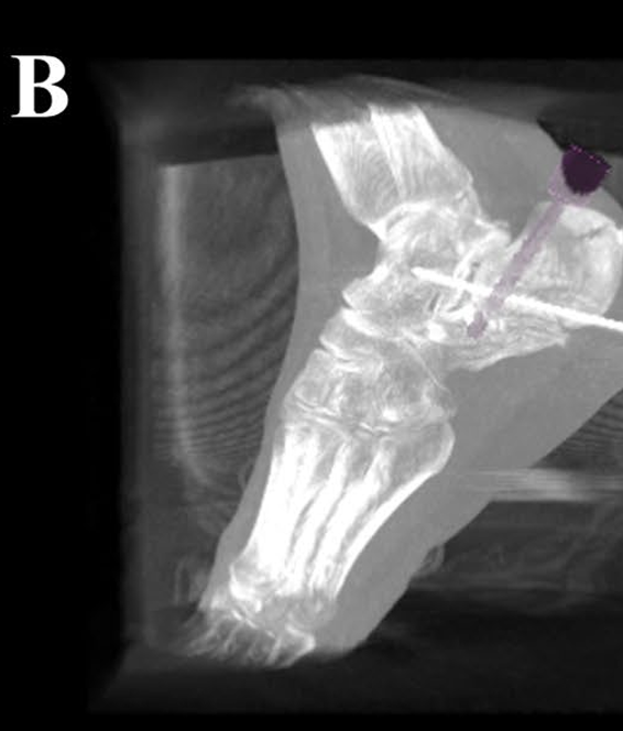
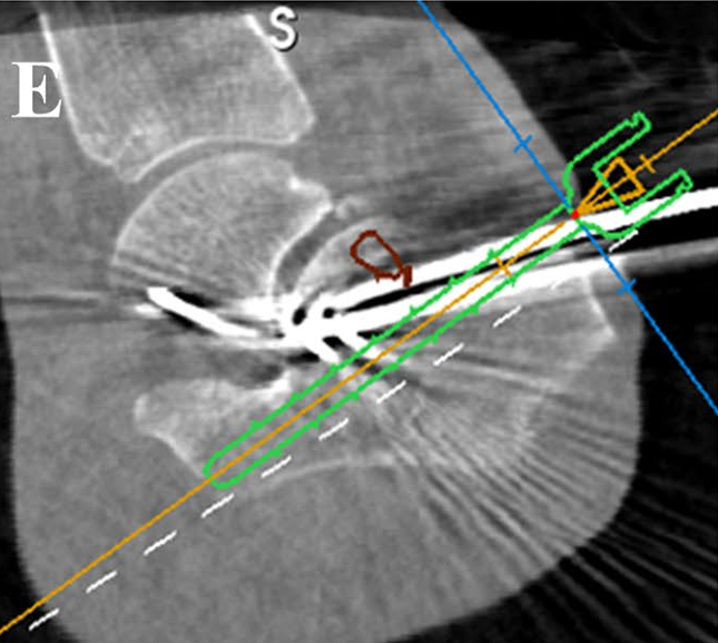
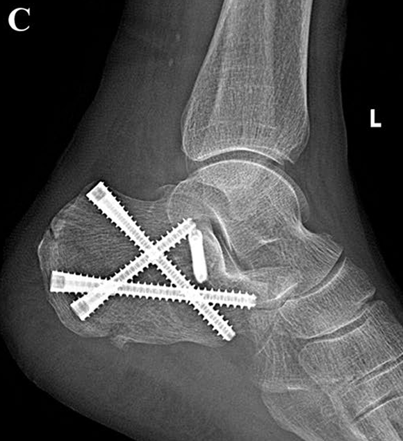
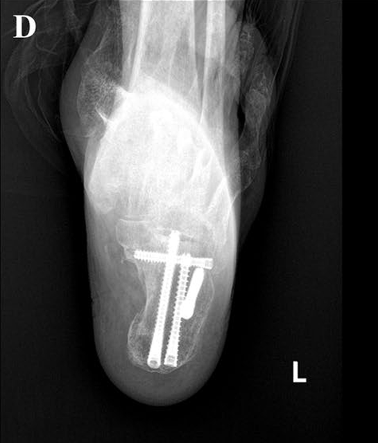
The patient was closely monitored postoperatively for signs of infection, pain management, mobility, and wound healing, considering her advanced age and medical history. Three days after surgery, Visual Analog Scale (VAS) score was reduced from 10 to 3. No complications were observed and the wound healed well. At 3 months, the patient had satisfactory clinical and functional outcomes with American Orthopaedic Foot and Ankle Society (AOFAS) score of 90. Follow-up X-ray shows the screw position is stable and unchanged (Fig. 3, C, D). This suggested the potential for good short-term results with this minimally invasive robot-assisted approach. However, longer follow-up is required to fully evaluate outcomes.
Discussion and Conclusion
The treatment of calcaneal fractures has advanced from conservative methods to minimally invasive surgical techniques that optimize subtalar joint and calcaneal integrity with fewer soft tissue complications [4, 5]. Minimally invasive surgery for calcaneal fractures have demonstrated significant improvements in clinical outcomes compared to traditional open reduction and internal fixation methods [6]. Tomensen et al. firstly introduced a method for treating displaced intra-articular calcaneal fractures using closed reduction and percutaneous screw fixation. The found that while the technique generally led to satisfactory functional outcomes and patient satisfaction, subsequent subtalar arthrodesis and the removal of irritating screws were sometimes necessary [7]. Long et al. presents a successful three-step closed reduction and percutaneous screw fixation protocol for displaced calcaneal fractures (even Sanders III and IV), evidenced by significant postoperative improvements and no reported complications [8].
Robot-assisted surgeries facilitate accurate fracture reduction with less radiation exposure and improved screw placement accuracy [9, 10]. The integration of robotics into the surgical treatment of calcaneal fractures has further enhanced the precision and safety of minimally invasive procedures. The use of robotic assistance has shown to significantly improve the function of the affected foot while maintaining the accuracy of screw implantation after fracture reduction, as demonstrated by Yuan et al. in their comparison of robot-assisted internal fixation and traditional methods for calcaneal fractures [11]. Wang et al. compares robot-assisted percutaneous screw fixation to ORIF for calcaneal fractures, concluding that the robot-assisted method offers a safe, effective, and less invasive alternative with improved postoperative outcomes and no significant complications [2]. The advantages of combining robotic technology with minimally invasive techniques are becoming increasingly apparent. This synergy allows for enhanced visualization, precision, and control during the surgical procedure, leading to improved anatomical restoration and reduced risk of postoperative complications. These benefits align with the principles of minimally invasive surgery – to achieve optimal clinical outcomes while minimizing tissue trauma.
The surgery in our case involved three critical steps for resolution. First, the two bone fragments had to be fixed together; simply repositioning the tongue-shaped bone fragment would not result in a satisfactory reduction of the articular surface. Second, traditional manual screw insertion is challenging when targeting particularly small bone fragments. Third, repeated attempts at K-wire insertion could lead to fragmentation of the bone pieces and decrease of biomechanical stability. For this case, the advantages of the robot were fully demonstrated. With robotic assistance, we achieved precise positioning of the two bone fragments, Kirschner wire insertion, and articular surface reduction in one attempt, followed by accurate screw placement. The total surgery time was 50 min, slightly higher than the average time of 48.6 ± 2.3 min reported by Yuan, which may be associated with the complexity of the surgery itself.
In addition to the points discussed above, we recognize the limitations of reporting a single case without a control group or longer-term follow-up. Nevertheless, this case highlights several clinically relevant aspects beyond serving solely as a technical note: (1) the unique patient context of an 85-year-old with a Sanders type III displaced intra-articular fracture and tongue-type morphology, where accurate reduction was critical to restore joint congruity; (2) the specific technical challenge in osteoporotic bone of achieving a single-pass Kirschner wire trajectory through two small, collapsed fragments without repeated attempts that could lead to bone fragmentation; and (3) the demonstrable role of robotics in enabling preoperative CT-based trajectory planning, precise single-attempt K-wire insertion, joystick-assisted fragment elevation, and optimized screw placement with minimal fluoroscopy exposure. These factors may not be reproducible with the same accuracy and soft-tissue preservation using freehand techniques. The patient achieved rapid pain relief, stable radiographic fixation, and good functional recovery at 3 months without complications, supporting the feasibility of this approach in selected elderly patients.
Looking forward, the future of robot-assisted minimally invasive treatment for calcaneal fractures appears promising. As technology continues to advance, we anticipate further enhancements in surgical precision, reduced operative times, and improved patient outcomes. Continuous research and clinical trials will be essential to establish standardized protocols and validate the long-term benefits of these innovative surgical approaches. Ultimately, the goal is to refine these methods to provide patient-centered care that optimizes function, minimizes complications, and accelerates recovery in individuals suffering from calcaneal fractures.
Declarations & Additional Information
Abbreviations:
CT: Computed tomography | MRI: Magnetic resonance imaging | VAS: Visual Analog Scale | AOFAS: American Orthopaedic Foot and Ankle Society | ORIF: Open reduction and internal fixation | MIS: Minimally invasive surgery
Acknowledgements: Not applicable.
Authors’ contributions: SL conceptualized the case report, drafted the initial manuscript, and contributed to the interpretation of data. CY F and XT L and XW analyzed the data, conducted the literature search, and assisted with writing and preparing figures for the manuscript. SY L served as a guarantor for the study, assisted in the surgical procedure, collected data, and participated in the manuscript review.
Funding: This work was supported by grants from the scientific research fund of Aerospace Center Hospital (no: YN202303), and Beijing Municipal Science and Technology Commission (no: Z221100007922003).
Data availability: No datasets were generated or analysed during the current study.
Ethics approval and consent to participate: Approval for the study by the local institutional review board was not required because it was a case report.
Consent for publication: Written informed consent was obtained from the patient for publication of this case report and any accompanying images.
Competing interests: The authors declare no competing interests.
Publisher’s note: Springer Nature remains neutral with regard to jurisdictional claims in published maps and institutional affiliations.
References
- Wu Z, Su Y, Chen W, Zhang Q, Liu Y, Li M, et al. Functional outcome of displaced intra-articular calcaneal fractures: a comparison between open reduction/internal fixation and a minimally invasive approach featured an anatomical plate and compression bolts. J Trauma Acute Care Surg. 2012;73(3):743–51.
- Wang J, Han W, Su Y, Wang J, Jiang X. Comparison of Robot-Assisted Percutaneous Cannulated Screws Versus Open Reduction and Internal Fixation in Calcaneal Fractures. Orthop Surg. 2023;15(3):724–30.
- Wang C, Xu C, Li M, Li H, Xiao H, Zhong D, et al. Patient-specific instrument-assisted minimally invasive internal fixation of calcaneal fracture for rapid and accurate execution of a preoperative plan: A retrospective study. BMC Musculoskelet Disord. 2020;21(1):407.
- Giannini S, Cadossi M, Mosca M, Tedesco G, Sambri A, Terrando S, et al. Minimally-invasive treatment of calcaneal fractures: A review of the literature and our experience. Injury. 2016;47(Suppl 4):S138–46.
- Wilkinson BG, Marsh JL. Minimally Invasive Treatment of Displaced Intra-Articular Calcaneal Fractures. Orthop Clin North Am. 2020;51(3):325–38.
- Wang Q, Chen W, Su Y, Pan J, Zhang Q, Peng A, et al. Minimally invasive treatment of calcaneal fracture by percutaneous leverage, anatomical plate, and compression bolts–the clinical evaluation of cohort of 156 patients. J Trauma. 2010;69(6):1515–22.
- Tomesen T, Biert J, Frölke JM. Treatment of Displaced Intra-Articular Calcaneal Fractures with Closed Reduction and Percutaneous Screw Fixation. J Bone Joint Surgery-American Volume. 2011;93(10):920–8.
- Long C, Li K, Zhu J, Liu H, Zhu Y. Three-step closed reduction and percutaneous screw fixation: A reliable and reproducible protocol in managing displaced intra-articular calcaneal fractures. Injury. 2023;54(Suppl 2):S49–55.
- Al-Naseem A, Sallam A, Gonnah A, Masoud O, Abd-El-Barr MM, Aleem IS. Robot-assisted versus conventional percutaneous sacroiliac screw fixation for posterior pelvic ring injuries: a systematic review and meta-analysis. Eur J Orthop Surg Traumatol. 2023;33(1):9–20.
- Bertelsen A, Melo J, Sánchez E, Borro D. Robot–assisted Percutaneous Pedicle Screw Placement Using Three–dimensional Fluoroscopy: A Preliminary Clinical Study. Int J Med Rob Comput Assist Surg. 2013;9(4):407–22.
- Yuan X, Tan K, Hu J, Zhang B, Zhang H. Does robot-assisted percutaneous hollow screw placement combined with tarsal sinus incision reduction in the treatment of calcaneal fracture perform better at a minimum two year follow-up compared with traditional surgical reduction and fixation? Int Orthop. 2023;47(6):1575–81.